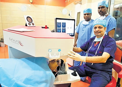

CHENNAI: Heads-up 3D visualisation is a technology used in various surgical procedures, including cataract and retina surgeries. It offers advantages over traditional surgical microscopes, such as improved depth perception and enhanced visualisation, improving surgical outcomes. It also enhances the ability to record and share surgical procedures for educational purposes. The state-of-the-art Zeiss Artevo 800 digital microscope is used in Rajan Eye Care Hospital to provide the best surgical outcomes for patients undergoing cataract and retinal surgeries.
Utility in Cataract Surgery
In cataract surgery, a 3D heads-up display allows surgeons to see a more detailed and three-dimensional view of the eye. This helps better identify the position and characteristics of the cataract and the surrounding eye structures. Depth perception is crucial in cataract surgery to safely remove the clouded lens and implant an intraocular lens. 3D visualisation aids in perceiving depth accurately which can lead to greater precision during surgery, potentially reducing the risk of complications. Surgeons can record surgeries and use the 3D heads-up display for teaching and training purposes, allowing newer surgeons to learn from experienced ones.
Utility in Vitreoretinal Surgery
Vitreoretinal surgeries often require delicate and precise manoeuvres on the thin and sensitive retina. 3D visualisation can provide a detailed view of the retina’s structure and pathology. Surgeons can magnify specific areas of the retina for close examination and perform microsurgery with greater accuracy. Procedures such as retinal detachment repair, diabetic vitrectomy, or macular hole closure benefit from the ability to precisely target and manipulate tissue using 3D visualisation. Recording and sharing 3D surgical footage is valuable for documenting cases and consulting with other specialists.
The equipment required for heads-up 3D visualisation typically includes a high-definition 3D display, a camera system mounted on the surgical microscope, and specialised software to process and display the 3-D images. Surgeons wear polarised glasses to view the 3D images on the display. The surgery can be simultaneously viewed by multiple people wearing polarised glasses in the operation theatre. While heads-up 3D visualisation requires specific training for surgeons to adapt to this technology. Overall, 3D heads-up visualisation has the potential to enhance the precision, safety, and educational aspects of cataract and retina surgeries, benefiting both surgeons and patients.
Robotic Cataract Surgery (CATALYS)
Cataracts are a common eye condition that can cause cloudy or blurry vision. Cataract surgery is a procedure to remove the cloudy lens of the eye and replace it with an artificial lens called an intraocular lens (IOL). This surgery is typically done to improve vision and reduce the impact of cataracts on daily life.
Femtosecond laser-assisted cataract surgery (FLACS) is an advanced and safe technique which utilises a femtosecond laser to perform the key steps in cataract surgery. It is a more precise and automated approach to traditional cataract surgery. It involves the use of a femtosecond laser, which emits extremely short pulses of light, to create incisions in the cornea, open the lens capsule, and soften the cataract for easier removal.
FLACS enables highly accurate and reproducible incisions, which can improve the visual outcomes. It provides consistent results, reducing the likelihood of variations in surgical technique and outcomes between different surgeons. It reduces the amount of ultrasound energy needed to break up and remove the cataract, potentially leading to quicker recovery and less inflammation. Astigmatism is a common refractive error of the eye that affects the way light enters the eye and is focused on the retina, resulting in distorted or blurred vision at various distances. FLACS can correct astigmatism more precisely than manual incisions. The laser can be customised to the patient’s unique eye anatomy, allowing for a tailored treatment approach that enhances the accuracy of the procedure. The use of a laser can reduce the risk of human error during certain steps of the surgery, potentially decreasing the risk of complications.
Prior to the surgery, your eye surgeon will examine you and determine the suitability for the procedure. Routine pre-operative workup similar to phacoemulsification surgery is done before the procedure to assess your individual eye health and recommend the most appropriate treatment plan for your specific needs. During the procedure, you will be positioned under the laser, and a computer-controlled laser will make precise incisions and perform other necessary steps. Once the laser application is completed, your surgeon will remove the softened cataract using ultrasonic technology and insert an IOL to replace the removed lens, which will typically be customised to your eye’s needs. This can be combined with monofocal IOL and all premium IOLs available in the market. The major advantage of FLACS is early post-operative recovery. While recovery time varies from person to person, some individuals experience quicker visual recovery after FLACS compared to traditional surgery. Studies also support the fact that FLACS may lead to reduced post-operative inflammation compared to traditional surgery, which can contribute to a more comfortable recovery eye drops and oral medications will be prescribed post-operatively similar to phacoemulsification. As with any surgery, there are potential risks and complications associated with FLACS, including infection, inflammation, bleeding, and changes in eye pressure.
Robotic cataract surgery offers the followimg merits
Safety, precision, accuracy, predictability, faster recovery
It is bladeless
Robotic cataract surgery is very dafe in hard brown cataracts, mature cataracts, postrior polar cataracts, cataracts with pseufoexfoliation, subluxated cataracta, Fuch’s edothelial dystrophy
(The writer is the chairman and medical director of Rajan Eye Care Hospital Pvt Ltd)